
Dental radiographs are diagnostic images produced when safe beams of electromagnetic energy pass through oral tissues onto a digital sensor. Anatomical structures absorb this energy based on physical density revealing what no clinical examination alone can uncover.
Digital radiography captures high-definition images instantly on a chairside screen, allowing your dentist to evaluate subtle details while reducing radiation exposure by up to ninety percent. The American Dental Association and the Food and Drug Administration recognize digital radiographs as essential tools for accurate patient care.
Enamel, dentin, and bone absorb substantial energy, appearing white or light gray on the image described clinically as radiopaque.
Gums, cheek tissues, nerves, and ligaments permit beams to pass through easily, appearing dark or black described as radiolucent.
When bacteria demineralize enamel or infections break down bone, the area loses density, creating a dark shadow inside the tooth structure.

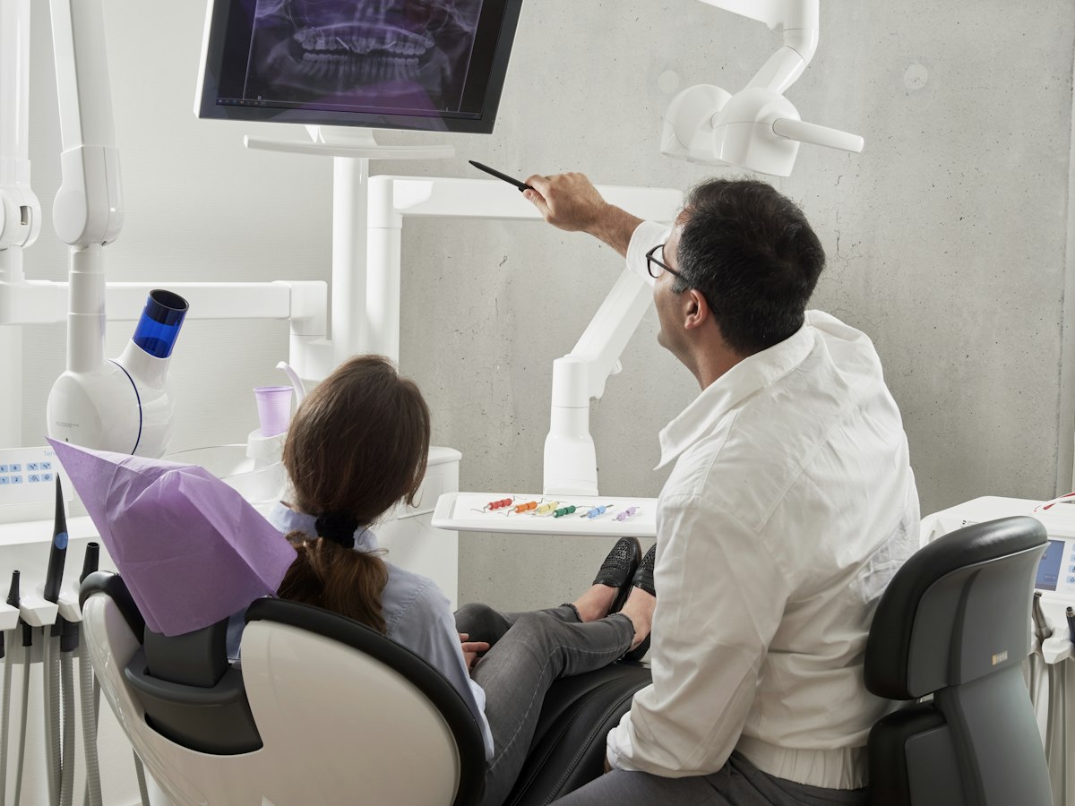
State-of-the-art digital radiography at Alani Dental — Jacksonville & Fleming Island, FL
Diagnostic radiography serves as an early warning system for your mouth — identifying conditions when they are small, manageable, and far easier to treat.
Interproximal decay forms where teeth touch and grows unnoticed for months. On a digital radiograph, decay appears as a dark notch in enamel — allowing your dentist to stop it with fluoride or a small composite filling before structural damage occurs.
When bacteria reach the pulp, infection travels down root canals into bone. Fluid and bacteria destroy bone around the root tip, forming an abscess. Radiographs reveal this bone loss, enabling treatment before infection spreads.
Periodontal disease attacks the underlying jawbone supporting teeth. Dental X-rays can provide information that may not be visible during a clinical examination.
Wisdom teeth frequently lack room to erupt normally, becoming impacted under gums or tilted against neighboring molars. Panoramic radiographs provide a complete view of the jaw to monitor wisdom teeth before complications arise.
Benign cysts, tumors, and abnormal bone growths can develop silently within jawbones without swelling or pain. Routine radiographs identify these anomalies during checkups, allowing for early specialist care.
Fillings, crowns, and bridges experience continuous chewing forces. Over time, margins can open, permitting bacteria to seep underneath. Radiographs are the only reliable way to evaluate tooth health beneath an intact restoration.
Dentists use specialized varieties of radiographs, each designed to capture specific perspectives and evaluate particular oral structures.
| Radiograph Type | Anatomical Focus | Primary Clinical Applications |
|---|---|---|
| Bitewing | Crowns of upper and lower teeth; crestal bone height |
Most common at checkups. Detects interproximal cavities and assesses bone levels between teeth.
|
| Periapical | Complete tooth from biting surface to root tip and surrounding bone |
Diagnoses root infections, assesses fractures, and monitors dental implants in detail.
|
| Panoramic | All teeth, jawbones, joints, and sinuses in a single wide-field scan |
Evaluates wisdom teeth development, overall bone health, and jaw anatomy at a glance.
|
| CBCT (3D Cone Beam) | Cross-sectional slices of teeth, jawbone, and nerve pathways |
CBCT may be indicated when detailed three-dimensional information is valuable for diagnosis or treatment planning.
|
Modern digital dental radiographs represent one of the lowest-dose diagnostic procedures in healthcare — and frequency is always personalized to your individual risk factors.
Expose you to approximately 0.005 mSv — less radiation than you absorb from natural background sources in a single day.
An individual absorbs roughly 0.008 mSv daily from the sun and earth — meaning 4 bitewings deliver less than a single day of natural exposure.
A commercial flight exposes you to 0.03 mSv of cosmic radiation — equal to six complete sets of bitewing radiographs.
A standard medical chest X-ray delivers about 0.1 mSv — equivalent to twenty full sets of dental bitewing radiographs.
Adults with frequent cavities, dry mouth, or periodontal disease require bitewings every 6 to 12 months. Children with active decay also require frequent imaging because cavities spread quickly through baby teeth.
Adults who brush and floss daily, maintain excellent hygiene, and have not developed cavities for years need bitewings every 12 to 24 months based on the ADA and FDA guidelines.
The type and frequency of dental imaging depend on the patient’s history, symptoms, clinical findings, and individual diagnostic needs.
Medical organizations confirm that necessary dental X-rays with lead apron shielding are safe throughout pregnancy. Untreated dental infections pose serious risks to maternal and fetal health — making prompt imaging essential when clinically indicated.